Make a commitment to your health with a 3D Mammogram
Clinical studies show 3D mammography provides better visualization of dense breast tissue and improves overall cancer detection rates.
To schedule online, see the details below or call for an appointment at
732-390-0033
To schedule an exam online you will need...
- Your Insurance Card
- Order / Script
- Name of your referring physician
- A contact email address or cell phone number
What is a 3D mammography breast exam?
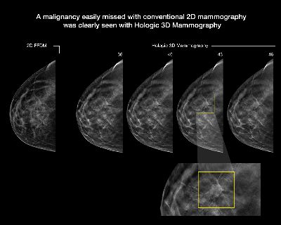
3D mammography is a new diagnostic tool that is used together with traditional 2D imaging to build a finely detailed 3D reconstruction of breast tissue. Radiology physicians can then scroll through all the images much like a book - one page at a time - to gain a better understanding of your breast health.
The 2D flat image on the far left indicates an area needing further study. 3D imaging of the same area with multiple slices from different angles provides additional detail.

What are the benefits of 3D mammography?
Sometimes breast tissue can overlap, which when viewed only with conventional 2D mammography, may look like an abnormal area. Examining multiple images with 2D and 3D techniques allows the radiologist to view tissue more clearly, making it easier to make an accurate assessment and less likely that you will be asked to return for additional views. Abnormalities may be easier to see and evaluate, making it possible to identify cancers at earlier stages when they are most treatable.
Is 3D mammography used for screening and for diagnostic exams?
A screening mammogram is a routine evaluation of the breast. A diagnostic mammogram focuses on an area of the breast that is unclear or is used to evaluate a symptom or complaint. Conventional 2D and the new 3D mammography may be used together to screen or to diagnose a breast issue.
What should I expect during my 3D mammography exam?
Traditional 2D and 3D mammography are performed on similar equipment. No additional compression or positioning is required during the 3D exam, and it only takes a few seconds longer than the 2D exam for each view.
During the 3D image acquisition, the X-ray arm will move in an arc over the breast, taking multiple images over the course of a few seconds. The technologist will view the images to ensure they are of the highest quality. The radiology physician will then study the images and dictate the findings in a report to send to your doctor. Your doctor will discuss the results with you.
Is there additional radiation exposure with 3D mammography?
With the more advanced, lower dose C-ViewTM software, the radiation used for the 2D together with the 3D exam is now about the same as with a traditional 2D mammogram alone.
Are there additional costs?
Some insurance companies do not yet cover 3D mammograms, but legislation and coverage are always changing. Please check with your insurance provider to understand your health care policy. There may be an additional out-of-pocket charge to you. For more information regarding insurance click on our insurance navigator below.
The American College of Radiology (ACR) and Society of Breast Imaging (SBI) recommend that women start getting annual mammograms at age 40. The American Cancer Society (ACS), US Preventive Services Task Force (USPSTF), ACR and SBI agree that this approach saves the most lives.