
Ultrasounds at University Radiology

Transabdominal vs. Transvaginal Ultrasounds | Preparation | Types of Ultrasounds | Women’s Health Sonograms | FAQs

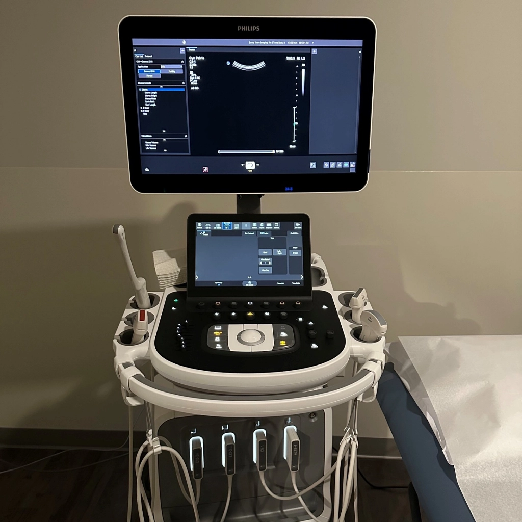
Ultrasonography is a method of examining parts of the body without the use of X-rays. Instead, it passes harmless sound waves through the body to record a picture on film. Ultrasounds can be used to assess pregnancy, diagnose certain medical conditions, and guide physicians during interventional radiology procedures.

Ultrasounds are extremely useful because they are portable, widely available, and radiation-free. They also cost less than other imaging studies, like CT scans. They are the preferred technique for collecting real-time images of fluid-filled organs and collections of fluid in other areas of the body.
ultrasound

Transabdominal vs. Transvaginal Ultrasounds

During a transabdominal ultrasound exam, a water-soluble “gel” is put on the skin over the area of concern. A sonographer will hold a transducer or probe (wand) and move it over the area to capture images. Transabdominal ultrasounds are completely noninvasive and usually painless.

In some women’s health cases, a transvaginal (TV) ultrasound is needed. Instead of an external probe, the sonographer or the patient inserts a slender probe into the vagina. The probe is covered with a sterile condom for the patient’s protection, as well as a water-soluble gel. The sonographer takes the end of the probe and gently moves it from side to side to get a clear view of the uterus, ovaries, and bladder. This exam should be painless, although patients with certain pelvic issues may feel some discomfort.

Preparing for Your Ultrasound

Preparation instructions for ultrasound examinations vary widely depending on the test ordered. For example, bladder sonograms require the patient to drink 24 ounces of water 1.5 hours before their test to fill the bladder. Pregnancy and pelvic ultrasounds also require drinking specific amounts of water at specific times. In contrast, patients getting abdominal sonograms must stop eating and drinking at midnight the night before their test.

All ultrasound exams require a written prescription or referral stating the exam being ordered and the patient’s signs and symptoms indicating medical necessity of the procedure. Patients should bring previous films or related exams to their appointment.

Common Types of Ultrasound Studies

At University Radiology, we offer a range of outpatient ultrasound procedures.

  • Abdominal Sonogram: Looks at the upper abdomen, including the gallbladder, liver, bile ducts, pancreas and spleen.
  • Aorta Sonogram: Checks for aortic aneurysm.
  • Bladder Sonogram: Evaluates the bladder, usually while it is full and/or just after urination.
  • Carotid Doppler Sonogram: Detects narrowing, blockages, and blood clots in the carotid arteries.
  • Elastography: Detects fibrosis (scar tissue) on the liver, which can lead to cirrhosis (liver disease).
  • Hip Sonogram: Checks for hip dysplasia and fluid buildup around the hip joint (effusion).
  • Neurosonogram: Assists in the diagnosis of hydrocephalus in infants.
  • Pelvic Sonogram: Assesses pregnancy, gynecological problems, abscesses, and pelvic or lower abdominal masses.
  • Renal Sonogram: Identifies masses, obstructions, or calcifications in the kidneys.
  • Scrotal Sonogram: Highlights masses in or inflammation of the scrotum.
  • Thyroid Sonogram: Detects thyroid masses or gland enlargement. Thyroid sonograms are often completed after nuclear medicine scans.

Women’s Health Sonograms

  • Breast Sonogram: Clarifies the nature of a breast mass seen on mammography or investigates a palpable lump. This exam should not be performed for patients with breast pain only.
  • Breast Aspiration: Removes fluid from a breast cyst.
  • Pregnancy Sonogram: Assesses pregnancy across many stages, including fetal anatomy, placenta placement, uterine health, etc.
  • Sonohysterogram: Examines the lining of the uterus (endometrium) to identify abnormal anatomical structure, detect fibroids and scarring, and investigate unusual bleeding. The procedure requires an injection of saline into the uterus.

Frequently Asked Questions

No, ultrasounds do not use radiation. Instead, they use sound waves to view structures in the body. Ultrasounds are not harmful to pregnant women.

Result times can vary based on the test ordered. After your scan, a radiologist reviews the images and sends a detailed report to your doctor. Once your doctor receives the results, they should reach out to you to discuss them.

While most ultrasounds are painless, certain conditions can make transabdominal and transvaginal ultrasounds uncomfortable. The sonographer may have to push the probe in different areas, which can agitate issues like hernias and ovarian cysts.

Most insurance providers cover ultrasound procedures. For more information and help understanding your coverage, call us at 732.390.0033.

Yes, sonogram and ultrasound refer to the same procedure.

Most ultrasound exams create 2D images on film. A 3D ultrasound uses multiple 2D images from different angles to create a 3D image. 3D ultrasounds are often used during pregnancy sonograms in the second and third trimesters.

Schedule an Ultrasound Near You

To schedule a consultation or ultrasound appointment with one of our specialists at a location near you, call 732.390.0033 or use our online scheduling system.